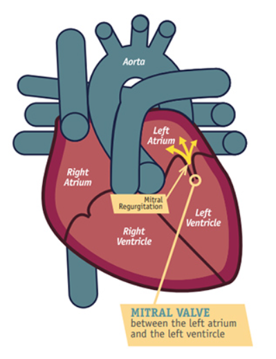
The Mitral Valve is a one-way valve that allows oxygenated blood (having arrived from the lungs into the left atrium) to flow from the left atrium into the left ventricle, ready to be pumped all around the body. Mitral regurgitation (MR) is a condition in which the mitral valve doesn’t close properly. When this happens, some blood leaks backwards with each heartbeat instead of moving forward into the body. Over time, this backward flow causes lung congestion and extra strain upon the heart, leading to breathlessness, fatigue, palpitations, or ankle swelling.
Mitral regurgitation can develop for several reasons, including natural wear and tear of the valve tissue, mitral valve prolapse, or conditions that affect the muscle or structures supporting the valve.
The seriousness of mitral regurgitation varies from person to person, as some people live with a mild leak for years while others may develop more significant symptoms that require treatment.
Diagnostic Methods For Mitral Regurgitation
Echocardiography (echo) is usually the first and most important test used to diagnose and assess mitral regurgitation. It allows us to see the valve moving in real time, measure the severity of the leak, and assess the impact on heart chambers and pressures. Regular echocardiograms are essential for tracking changes over time and guiding decisions about when treatment (whether medication or surgery)might be needed. The rate at which mitral regurgitation progresses varies widely, which is why regular scans are used to see whether anything has changed.
In many patients, additional imaging can provide valuable detail. Cardiac MRI is the gold-standard method for precisely measuring heart chamber size and function, and it offers highly accurate quantification of the degree of regurgitation. This can be particularly helpful when echocardiographic measurements are unclear or when we need a more comprehensive understanding of how the condition is affecting the heart.
Cardiac CT can also play an important role, especially when evaluating the anatomy of the mitral valve and surrounding structures before procedures such as mitral valve repair or replacement, but also in assessing the coronaries for any underlying coronary disease, which may sometimes be the cause, thereby requiring its own intervention. CT offers excellent spatial detail and helps plan interventions safely and effectively.
Some people experience no symptoms for many years, which is why careful monitoring is important if it is detected early. If it is severe, treatment is initially with tablets to offload the congestion from the lung, which is often then followed by an operation to repair or replace the mitral valve.
If you are concerned about mitral regurgitation or would like to discuss anything above, please use our appointments page to book a consultation.
